![]()
Advanced OCT
# A faster, better way of seeing
A Faster, Better Way of Seeing
NinePoint Medical’s NvisionVLE® Imaging System with Real-time Targeting&trade is a groundbreaking convergence of superior optical imaging, innovative engineering techniques, and revolutionary information technology. At the heart of the system, Advanced Optical Coherence Tomography (OCT) technology provides physicians and pathologists with comprehensive, volumetric digital images of a patient’s esophagus. This next-generation technique enables much higher imaging speeds, combined with improved field-of-view and signal-to-noise ratio.
For resources on OCT technology click here.
How Advanced OCT Works
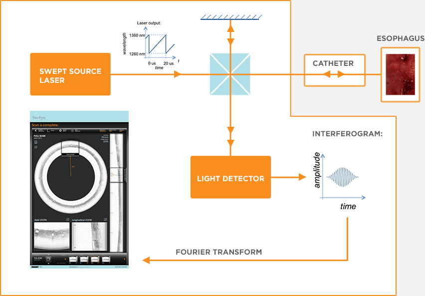 The (greatly simplified) fundamentals of Advanced OCT are shown in the diagram. Similar to a traditional laser, the swept-source laser produces light of only one ‘color’ at any instant in time. However, unlike a traditional laser, the swept-source changes the color of its output light very rapidly in time.
The output light is split in two: one beam stays internal to the console, while the second is transmitted through the catheter and focused into the tissue. Some of the light is reflected back from physiological structures within the tissue, and this reflected light is collected by the catheter and returned to the console.
Inside the console, the reflected and internal beams are combined and interfere with each other. After some processing, the interference signal yields an axial line (‘A-line’) that records the intensity of reflections at each depth in the tissue. Combining many consecutive A-lines into an image provides a cross-sectional look at all the reflecting structures in the tissue.
A Brief History of OCT

 The basic principles of Optical Coherence Tomography (OCT) were initially used within the fields of optical radar and optical telecommunications, among others. The technology expanded in the late 1980s when researchers began exploring applications for biological tissue imaging. Over the next decade, researchers around the world developed sub-categories of the technology, such as swept-source OCT, and began investigating ways to use OCT in different types of tissues and biomedical applications.
Today, commercial OCT systems are used regularly as an imaging standard in ophthalmology, cardiology, dermatology, and general research. NinePoint’s Advanced OCT technology, licensed from the Wellman Center for Photomedicine at Massachusetts General Hospital, initially focuses on esophageal and general imaging of tissue microstructure.
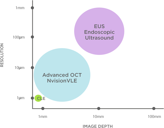 Filling an Imaging Gap
The ideal imaging system for esophageal surveillance would combine high-resolution scanning, depth, and volume to enable the identification and interrogation of both superficial and buried features.