![]()
NvisionVLE Imaging System
# with Real-time Targeting™
 The NvisionVLE® Imaging System, with Real-time Targeting™, as part of the VLE procedure (Volumetric Laser Endomicroscopy), uses an optical signal acquisition and processing method to create high-resolution cross-sectional images and mark tissues—letting you evaluate 100% of the tissue in a 6cm scan in real-time. 3mm deep. At a resolution of 7 microns. All so you can provide a more thorough evaluation – potentially leading to improved biopsy targeting for diagnosis by histopathology, and more complete information to determine the best treatment for your patients.
The NvisionVLE Imaging System is indicated for use as an imaging tool in the evaluation of human tissue microstructure, including esophageal tissue microstructure, by providing two-dimensional, cross-sectional, real-time depth visualization and may be used to mark areas of tissue. The safety and effectiveness of this device for diagnostic analysis (i.e. differentiating normal versus specific abnormalities) in any tissue microstructure or specific disease has not been evaluated.
A Streamlined Procedure
AThe NvisionVLE® Imaging System with Real-time Targeting™ allows clinicians to evaluate the esophageal tissue microstructure and mark areas of suspicion that are not visible with conventional imaging modalities, during a standard endoscopy procedure. These features help provide a more thorough evaluation—potentially leading to improved biopsy targeting for diagnosis and more effective therapeutic decisions for patients.
NvisionVLE® Imaging System with Real-time Targeting™ Animation
An Advanced System
 NvisionVLE® Imaging System with Real-time Targeting™ Advantages:
- Uses Advanced OCT to capture images up to 3mm beneath the mucosa at a 7 micron resolution in real time—unlike white light endoscopy, which can only image surface detail
- Offers a volumetric view (~10,000mm2), as opposed to a “point” image typically obtained with confocal microscopy (0.25mm2)
- Advanced Optical Coherence Tomography (OCT) imaging delivers up to 25X higher resolution than endoscopic ultrasound
- Provides a dramatic increase in imaging speed and improved image resolution, compared to first-generation OCT systems
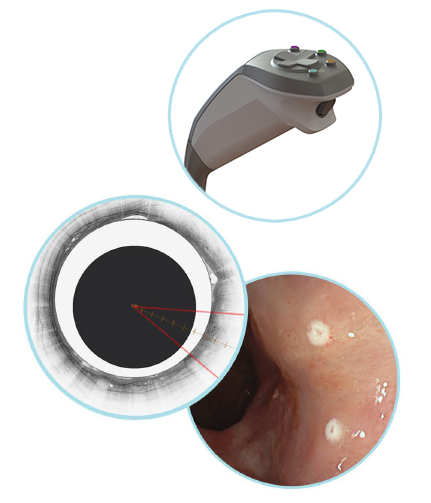
- Creates tissue laser marks visible under white light endoscopy, designed to help clinicians target biopsies at a site of interest that may not be visible with other imaging modalities
A Closer Look
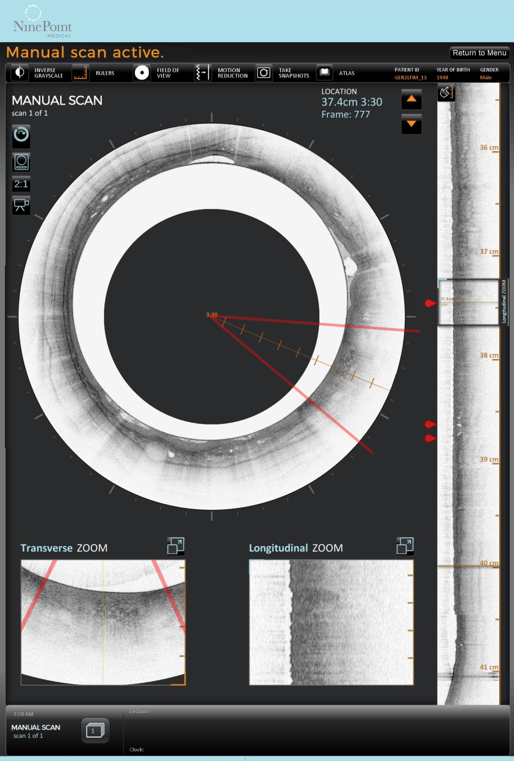 Cross-Sectional View
Explore the entire targeted segment of the esophagus in real time:
- Collect 1,200 cross-sectional images
- Across a targeted segment of 6cm
- Penetrating approximately 3mm into esophageal tissue
For close examination of a particular area of interest, either within the cross-sectional or longitudinal views, these windows provide a zoomed-in view.
Longitudinal View
Examine the plane of the esophagus perpendicular to its cross-section:
- View the esophageal wall along the axis of the organ
- With over 4,000 longitudinal images of the esophagus
All Views
As you manipulate the cross-sectional or longitudinal views on the NvisionVLE touch-screen monitor or hand-controller, each of the corresponding views update smoothly and in real time.
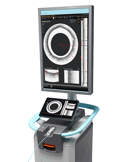
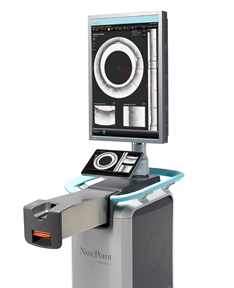 Image Console & User Interface
The NvisionVLE® Imaging Console is designed with clinicians in mind:
- Hand-controller allows for real-time physician control of the VLE scan and tissue marking to better evaluate areas of concern
- Active clinican-controlled workflow, tailored to the specific needs of each patient
- Single and double mark options for application flexibility
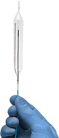 A High-Speed Optical Probe
- Balloon catheter facilitates optical probe positioning and centering (available in 14mm, 17mm and 20mm sizes)
- Also available as balloon-less 7 French Low-Profile optical probe to accommodate various anatomies
- Catheter compatible with endoscope channels 2.8mm or larger
Reimbursement
The Current Procedural Terminology (CPT)* codes used for reimbursement of the VLE (Volumetric Laser Endomicroscopy) procedure are:
- CPT 43206 Esophagoscopy, flexible, transoral; optical endomicroscopy
- CPT 43252 Esophagogastroduodenoscopy, flexible, transoral; optical endomicroscopy
- CPT 0397T – Endoscopic retrograde cholangiopancreatography with optical endomicroscopy
- CPT 88375 Optical endomicroscopic image(s), interpretation and report, real-time or referred, each endoscopic session
To receive the NvisionVLE® Imaging System Reimbursement Guide, please request and complete this form or ask your local NinePoint Medical representative.
For more information about reimbursement, please contact us at reimbursement@ninepointmedical.com.
* CPT codes and descriptions only are copyright 2017 American Medical Association (AMA). All rights reserved. The AMA assumes no liability for data contained or not contained herein.
The coding, coverage, and payment information contained herein is gathered from various resources and is subject to change without notice. NinePoint Medical cannot guarantee success in obtaining third-party insurance payments. Third-party payment for medical products and services is affected by numerous factors. It is always the provider’s responsibility to determine and submit appropriate codes, charges, and modifiers for services that are rendered. Providers should contact their third-party payers for specific information on their coding, coverage, and payment policies.